In this training video linda bevington bsn rn cpn patient family educator at chop demonstrates the equipment needed and the steps to take before placin. The end of the tubing left o.
 Nasogastric Ng Tube Insertion Osce Exam Demonstration Youtube
Your nurse will tell you the best way to hold your child.

Nasogastric tube insertion video. A nasogastric tube ng tube is a fine tube that is passed through your nose down the back ofyour throat and into your stomach. We need to lubricate the tube because we want it to slide down the nasal passage without causing any irritation. In this training video linda bevington bsn rn cpn patient family educator at chop demonstrates how to position and secure your child for ng tube insert.
This process is known as nasogastric ng intubation. This involves inserting a tube through the nasal passage into the stomach. Nasogastric ng feeding tube insertion training video.
Dip the tube in the lubricant. Creative commons licensing attribution non commercial. About press copyright contact us creators advertise developers terms privacy policy safety how youtube works test new features press copyright contact us creators.
Simple steps for inserting an ng tube. Now it is time to put the tube in. By inserting a nasogastric tube you are gaining access to the stomach and its contents.
What is the normal potassium level. Potassium normal range htq potassium normal range a normal range for potassium is between 3 6 and 5 2 millimoles per liter mmol l of blood.
 Jpma Journal Of Pakistan Medical Association
If potassium level in blood is between 5 0 to 6 0 mmol l it is called mild hyperkalemia.
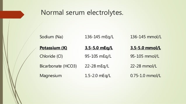
Normal range for potassium in blood. Potassium is an essential ion found within the cells of the body. 5 4 potassium level i had blood test for surgery and all were normal range except k how can i lower my potassium answered by dr. The normal range for potassium in the blood is between 3 6 and 5 2 millimoles per liter according to healthline.
Your doctor may want you to get a blood test. A typical potassium level for an adult falls between 3 5 and 5 0 millimoles per liter mmol l. What is the normal potassium range.
The normal range of potassium in your blood is between 3 5 to 5 0 millimoles per liter. Although the normal level of potassium in the blood varies in different sources its average amount usually ranges from 3 5 to 5 0 meq l. Your body should maintain a specific amount of potassium in the blood ranging from 3 6 to 5 2 millimoles per liter mmol l.
Why would i get this test. On whether the high potassium level is because of kidney d. Hyperkalemia occurs when levels go above 5 5 mmol l.
The delicate balance between potassium outside the cell known as the extracellular fluid k and the potassium inside the cell called the intracellular fluid k helps maintain the electrophysiology of the body. High potassium levels hyperkalemia may be seen in conditions such as. A reading above 6 5 mmol l can cause heart problems that require immediate medical attention.
This sometimes varies depending on the laboratory that performs the test but the doctor uses the range marked on the results to make informed recommendations. This normal range also depends on some factors especially the age of the individual since the known normal range for children is in between 3 4 to 4 7 meq l. What is a safe or normal potassium level.
Hemoglobin is the main component of red blood cells. Normal ranges for children vary with age and sex.
 Hemoglobin Affinity For Oxygen In Three Subspecies Of Toads Bufo Sp Living At Different Altitudes
Hemoglobin is made up of four protein molecules globulin chains that are connected together.
Lab results hemoglobin. In the blood it carries oxygen to the cells in the body from the lungs. Hb is a red substance made of iron and protein. A low level of hemoglobin is usually associated with a disease or condition that results in a lack of red blood cells in the body.
For men 13 5 to 17 5 grams per deciliter. Normal ranges for hemoglobin in g dl. Lower than normal results.
For women 12 0 to 15 5 grams per deciliter. Children to up 18 years of age. The range for a normal hemoglobin level may differ from one medical practice to another.
The normal adult hemoglobin abbreviated hgb or hb molecule contains two alpha globulin chains and two beta globulin chains. This can happen if. The hemoglobin test is often used to check for anemia usually along with a hematocrit or as part of a complete blood count cbc.
The normal range for hemoglobin is. Hemoglobin is the protein molecule in red blood cells that carries oxygen from the lungs to the body s tissues and returns carbon dioxide from the tissues back to the lungs. The body produces less red blood cells than required aplastic anemia iron deficiency anemia cancer cirrhosis chronic kidney disease some medications.
5 or 20. If your hemoglobin level is lower than normal you have anemia. The test may be used to screen for diagnose or monitor a number of conditions and diseases that affect red blood cells rbcs and or the amount of hemoglobin in blood.
Create your own short introduction. Furthermore at times the medical reports are also wanted by the courts when the patient is seeking for an injury or accident compensation.
 Deciphering Your Lab Report Lab Tests Online
As for the students passing the tests and examinations in the laboratory is the main requirement for their curriculum.
Medical lab report. Learn about what a cbc is. One of the most common blood test analyzed in a medical lab is a complete blood count cbc. When i say writing that includes the careful analysis of data and attention to the details of how the information is formatted for the ultimate reader.
A cbc measures the most common types of cells and elements in your blood such as red blood cells rbc white blood cells wbc and platelets. If your professor requires you to conform to a specific. Every hospital has to prepare a medical report on every patient for further assessment of the patient s condition.
The format of references varies amongst journals. Tests may be run in a physician office laboratory a laboratory located in a clinic or hospital and or samples may be sent to a reference laboratory for analysis. Do not copy from the laboratory write ups.
Medical reports include the findings of the clinical examination conducted on a patient. In addition laboratories are considered as one of the main training evaluation grounds for the students especially those who are enrolled in medical courses. 13 50 0321 submitted to the department of science laboratory technology school of science and technology moshood abiola polytechnic pmb 2210 abeokuta ogun state in partial fulfilment of.
Many of your science units will require you to write formal laboratory reports. Technical report on student industrial work experience scheme siwes undertaken at orile agege general hospital o a g h 93 old ota road orile agege lagos state by utubor lucky osamudiame matric no. This is the date this copy of the report was printed.
Review the components of the science laboratory report. For your chemistry laboratory reports you should follow by default the acs guidelines as outlined in the acs style guide and journal of the american chemical society jacs all examples given in this handout conform to jacs format. Rbcs contain hemoglobin which carries oxygen to all your cells whereas wbcs are part of your immune system and help to destroy.
The purpose is to report on what you did what you learned from an experiment and why the findings matter. Select the report section that relates to the statement. Sample incident report forms.
View laboratory report sheet cell docx from bs bio 3102 at university of the city of manila pamantasan ng lungsod ng maynila. Name and address of the laboratory location where the test was performed. Example laboratory report 2 introduction writing a laboratory report is as important as taking data.
Medical forms in pdf.
Psychological adaptation the ongoing process anchored in the emotions and intellect by which humans sustain a balance in their mental and emotional states of being and in their interactions with their social and cultural environments. In biology has several meanings.
 Power Point Adaptations
When man first trekked out of africa into novel environments our physiology adapted so we could survive in novel conditions.
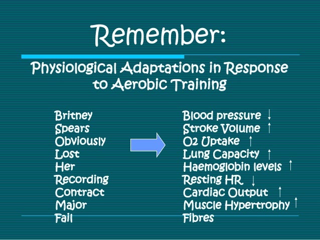
What are physiological adaptations. The thorny devil couldn t survive with just structural adaptations and instincts they need the chemical and internal adaptations physiological adaptations. Physiological adaptations in response to training physiological adaptations are always specific to the training and stress placed upon the body. Physiological adaptations internal and or cellular features of an organism that enable them to survive in their environment e g.
The word can also refer to a trait that is considered an adaptation. Physiological adaptation the ongoing process by which internal body functions are regulated and adjusted to maintain homeostasis in the internal environment. The human body lives and dies on its physiology and how it can adapt to novel environments.
Snakes produce poisonous venom to ward off predators and to capture prey. Humans have survived on this planet for thousands of years because of our ability to adapt. Please enable javascript and refresh the page to continue.
Physiological adaptations start to occur almost immediately when beginning a new exercise program. It can mean the adjustment of living matter to environmental conditions and to other living things either in an organism s lifetime physiological adaptation or in a population over many many generations evolutionary adaptation. Most animals physiologically adapt by developing means for protection body temperature regulation and predation.
Mammals that do frequent deep sea diving have a protein called myoglobin which stores oxygen in their muscles thus allowing them to dive for longer and enabling them to have a flexible rib cag. There are many physiological adaptations in animals. It is the adaptations that occur that cause the improvement in performance after training.
The bbc states that an animal can physiologically adapt to become tolerant to aridity chemical pollution cold temperatures hot temperatures altitude and fire. Training that uses the principles of training will cause more adaptations than training that does not. An adaptation that involves chemicals or chemical reactions.
Physiological adaptations to arctic climates the human body is one of the most amazing and complex biological systems on earth. Many changes occur throughout the body but the most significant changes include changes in the muscles bones and cardiovascular system.
What does a standard catheter kit include. Drape sheet with appropriate cutout.
 Amazon Com Bihiki Incontinence Kit Urinary Catheter Bag Ostomy Bag Holder Bladder Ostomy Elderly Drainage Bag Care Package With Adjustable Shoulder Strap For Home Travel Wheelchair Bed 1000ml 1500ml Health Personal Care
99 15 99 count 17 47 17 47.

Home catheter kit. The foley catheter is used to drain urine out from the bladder. Culture bottle for testing. Whether you need a specialized coude catheter or a long term solution with an intermittent catheter southwest medical offers several options from your trusted brands to meet your patient s needs.
Complete kit urinary incontinence one week 7 condom catheters external self seal 32mm intermediate premium leg bag 1000ml tubing straps fast and easy draining. A catheter insertion kit comes complete with everything necessary to perform the procedure as smoothly and sanitarily as possible. Reduce the risk of infection and cross contamination.
Available in 14 5fr diameter these tunneled polyurethane dialysis catheter kits come in a range of lengths and kit configurations that allow high flow rates and decrease patient therapy times. Usually a catheter kit includes the following items. Used to collect and store urine.
3 8 out of 5 stars 72 21 99 21. 1 csr wrap 1 catheter 1 tray 1 100 ml collection burette 1 pvp iodine swabsticks 1 lubricating jelly 5 2 x 2 gauze 1 paper poly towel and 1 drape. Complete kit urinary incontinence one week 7 condom catheters self seal external 29mm medium premium leg bag 1000ml tubing straps fast and easy draining.
Foley catheter kit we are the dealers of foley balloon catheterisation tray cathsafe urinary catheterisation tray foley insertion tray urinary catheter kit. Unecath newtech medical device s offers unecath catheter kit. It comes in single double lumen triple lumen with kink resistive catheter.
It stays in the bladder for a long period with the help of a retention balloon and is therefore called. Catheter kit 1 indwelling insertion tray 1 intermittent tray 1 intermittentent catheter kit 1 urinary catheter care kit 3 closed system catheter 2 urine meter tray 2 closed system intermittent catheter kit 1 groshong picc basic kit 1 indwelling catheter set 1 intermiitent catheter kit 1 intermittent catheter 1. Components of foley insertion kits and trays.
Foley insertion trays which come along with catheters generally have a foley catheter pre attached to a drainage bag. Our foley catheter kit includes. Can be an intermittent or foley catheter that is inserted into the bladder to drain urine.
Urinary drainage kits contain. Unecath covers almost every size in pediatric and adult length. The taurus dialysis catheter is indicated for use in attaining long term vascular access for hemodialysis therapy.
3 9 out of 5 stars 586 15 99 15.
It covers the latest technologies and methods for measuring cardiac output using biomedical signals monitoring pressure and flow and more. The one of a kind book provides comprehensive information on hemodynamic monitoring in a reader friendly style that s easy to understand.
 A Review Of Hemodynamic Monitoring Techniques Methods And Devices For The Emergency Physician The American Journal Of Emergency Medicine
Easy to compress to affect haemostasis following removal of the arterial line.
Hemodynamic monitoring made easy. Discuss the indications for invasive hemodynamic monitoring. Hemodynamic monitoring refers to measurement of pressure flow and oxygenation of blood within the cardiovascular system. Delineate hemodynamic values for pulmonary artery catheter arterial line and central venous pressure monitoring.
2005 real world nursing survival guide hemodynamic monitoring elsevier science united states of america nursing responsibilities during the insertion procedure include. Michael r pinsky 0 0 0 professor of critical care medicine bioengineering and anesthesiology university of pittsburgh medical center 606 scaife hall 3550 terrace street pittsburgh pa 15261 usa the book is divided into three sections. Compare preload afterload and contractility when determining cardiac function.
Assist with patient positioning and explanation to the patient. Describe three steps to ensure waveform accuracy. Or using invasive technology to provide quantitative information about vascular capacity blood volume pump effectiveness and tissue perfusion.
With this solid foundation of knowledge and current evidence based thinking readers will understand exactly what. Hemodynamic monitoring made easy critical care book report hemodynamic monitoring made easy michael r pinsky corresponding author.
It s common for an ostomy and pouch to go undetected. The quantity of ostomy supplies needed by a beneficiary is determined primarily by the type of ostomy its location its construction and the condition of the skin surface surrounding the stoma.

Colostomy care is how to change empty or clean your pouch system.

How to care for colostomy. Urine will automatically flow from the bag when it is opened. You and your family will be taught colostomy care before you leave the hospital. To prepare for irrigation the colostomy patient should take a seat on the toilet.
You and your family will be taught colostomy care before you leave the hospital. If necessary use a gloved and lubricated finger to dilate the stoma. If this is not possible use.
A colostomy can be temporary or permanent depending on the reason for its creation. If you have a descending or sigmoid colostomy you may choose to manage your colostomy with irrigation. Irrigation is simply putting water into the colon through the stoma to help regulate bowel movements.
For feces you can gently squeeze it out of the bag. The best place to do this is in the bathroom. Insert a flexible catheter coated in water soluble jelly no more than 3 inches 7 6 cm into the stoma.
A colostomy is a surgically created opening in the abdomen where a portion of the colon is brought through to allow feces stool to pass. Your healthcare team will help you do this. What you need to know.
Put on medical gloves. How do i empty my pouch. Colostomy irrigation has been used for many years but it s not used as much now as years ago.
Open the bottom of the bag over the toilet. A temporary colostomy will allow the affected bowel a chance to rest and heal. Promotes better evacuation of stool and avoids wrinkles on the colostomy.
1 x research source if there is urine or feces in your colostomy bag it is important to empty these prior to changing the bag. This allows you to meet and talk with people who understand what you re going through. Avoid drinks with caffeine or alcohol as these can irritate your digestive system after a colostomy.
Learning to care for and trouble shoot your colostomy will take some time. What is colostomy care. Step 1 start by emptying your colostomy bag.
Empty the pouch and remove the ostomy skin barrier. Unfasten belts if clients wear one. Empty the pouch when it is to full and before you change the system.
And look for simple non carbonated drinks like water and decaffeinated tea and coffee. Always empty the pouch through the bottom to prevent spillage of contents into the client s skin. Stop at the first sign of resistance.
Colostomy care is how to change empty or clean your pouch system. Alternatively some step 2 wash your hands thoroughly using soap and water. You may also want to try an ostomy support group.
Document if the diversion is an intestinal or urinary ostomy whether it s temporary or permanent and the location abdominal quadrant skinfold.
Alpha 2 adrenoceptors on the other hand are autoreceptors involved in the regulation of noradrenaline norepinephrine release. Whereas beta receptors can cause relaxation and dilatation.
 Alpha Blocker Wikipedia
Alpha and beta receptors are situated at the postsynaptic membrane of the sympathetic neuroeffector junctions of different organs.
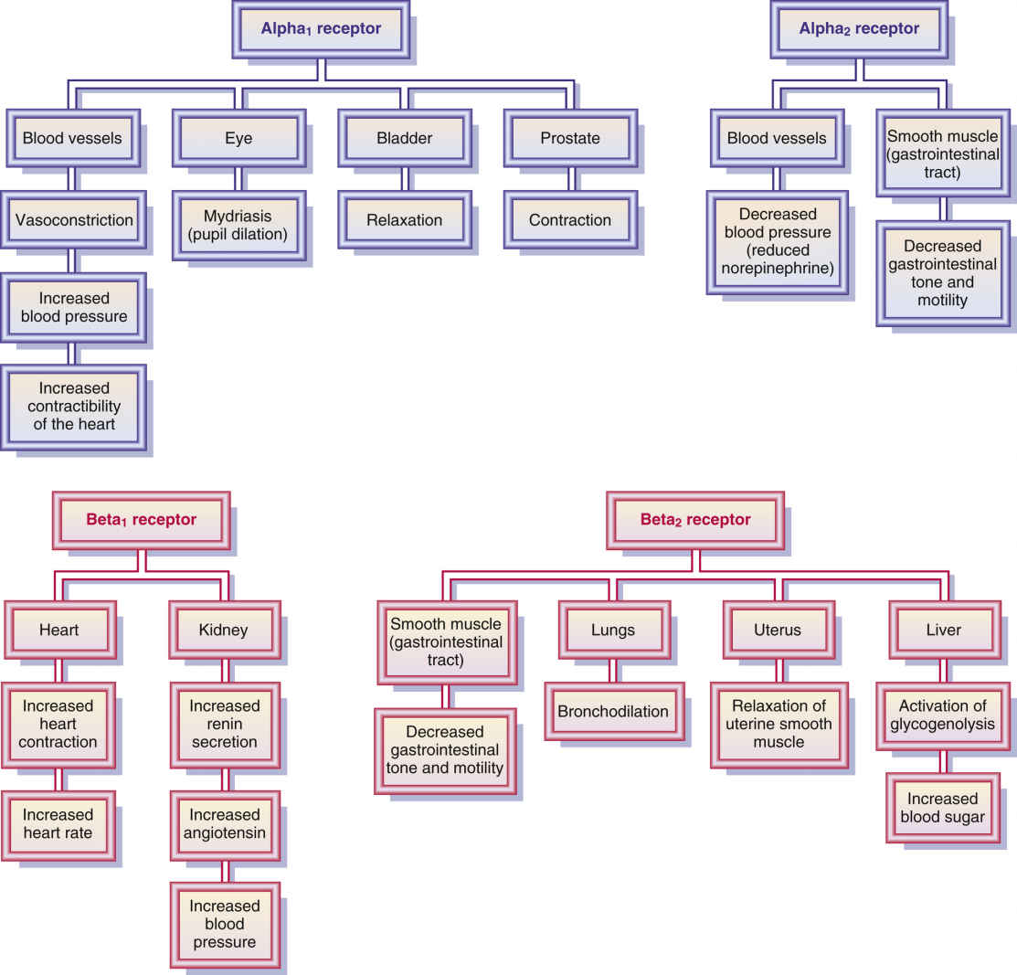
Alpha and beta receptors for dummies. Useful generalizations concerning these are. Alpha receptors can cause stimulation and constriction. Both alpha and beta receptors are innervated by the sympathetic nervous system.
Beta adrenergic receptors are one of two main adrenergic receptors the other being alpha receptors. This human anatomy and physiology video teaches functions of alpha vs beta adrenergic receptors. There are two categories of receptors alpha and beta.
Activation of alpha 1 and beta 1 receptors cause stimulatory responses. Specifically alpha receptors are found in arteries effector tissues and vascular smooth muscles while beta receptors are mainly located in involuntary muscles such as uterine bronchi and cardiac muscles. Both alpha and beta receptors are stimulated by epinephrine and norepinephrine.
Activation of alpha 2 beta 2 beta 3 receptors cause inhibitory responses. Both alpha and beta receptors are located on the postsynaptic membrane at the sympathetic junctions. These body processes become our local responses to particular stressors when we are faced with the fight and flight phenomenon.
Always remember you have one heart two lungs. Alpha is the excess return on an investment after adjusting for market related volatility and random fluctuations. Alpha receptors the alpha receptors or alpha adrenoceptors are those that respond to epinephrine and norepinephrine.
Both alpha and beta receptors stimulate or relax effector cells of several organs in the body. Ne causes a greater response than e when activating alpha 1 receptors. Alpha and beta receptor drug properties are discussed within this video.
Both norepinephrine and epinephrine can act on beta receptors however epinephrine generally has a higher affinity. They are involved in generating a sympathetic response when activated by catecholamines such as norepinephrine or epinephrine. In the periphery alpha 1 receptors are located postsynaptically mediating the excitatory effects of catecholamines at alpha receptors.
There are also subtypes of each. The adrenergic receptors are divided into two types. Alpha always constricts beta always.
Beta is a measure of volatility relative to a benchmark such as the s p 500. Difference between alpha and beta receptors. Subtypes of both alpha and beta adrenoceptors exist.
There are several types of alpha and beta receptors.
The 3m littmann classic iii stethoscope offers high acoustic sensitivity for exceptional performance when doing general physical assessments. 3m littmann master cardiology stethoscope 2182 smoke finishchestpiece and eartubes dark olive green tube.
3m Blacktube Smoke Finish Chestpiece Champagne Stem Cardiology Iv 6204 3m Littmann Stethoscope Shopee Singapore
Littmann stethoscopes offer superb acoustic performance consistently high quality and outstanding value.

Littmann smoke finish. Explore stethoscopes for adult and pediatric patients. Littmann cardiology stethoscopes are useful for diagnosing in critical care and challenging environments. Littmann cardiology iv stethoscope red smoke heart 6182.
Wearing a littmann stethoscope is an expression of your commitment to medicine and personal success. With a tunable diaphragm dual lumen tubing and a precision ergonomically designed stainless. It features dual tunable diaphragms and an updated design that is easier to clean and maintain.
3m littmann cardiology iv stethoscope smoke finish chestpiece turquoise tube smoke stem and headset 27 inch 6171. 3m littmann master cardiology stethoscope smoke finish chestpiece black tube 27 inch 2176. 3m littmann classic iii monitoring stethoscope smoke finish black tube 27 inch 5811.
3m littmann classic iii monitoring stethoscope 5811 smoke finish black tube 27 inch. The 3m littmann master cardiology stethoscope is characterized by the best acoustic response in the littmann mechanical stethoscope line. 3m littmann cardiology iv stethoscope high polish smoke chestpiece carribean blue tube and mirror.
The littmann cardiology iv diagnostic stethoscope model 6182 features a smoke finish chestpiece and binaurals with red tubing to create a beautiful look. At 27 in total length this model fits comfortably around your neck and stays. 3m littmann classic iii monitoring stethoscope 5871 smoke finish chestpiece black stem and headset raspberry tube catalog number 5871 upc 00707387783105 overview.
High polish smoke chestpiece carribean blue tube and mirror stem 6234.
Filter by location to see nurse anesthetist salaries in your area. We ve identified six states where the typical salary for a nurse anesthetist job is above the national average.

Bureau of labor statistics making it the top paying nursing specialty.

What is the average salary for a nurse anesthetist. Visit payscale to research nurse anesthetist crna salaries by city experience skill employer and more. The average salary for a nurse anesthetist is 150 111. Salary estimates are based on 10 salaries submitted anonymously to glassdoor by nurse anesthetist employees.
The estimate will vary depending on where you work and the state where you are employed. According to 2019 data from the bureau of labor statistics nurse anesthetists earn an average salary of 181 040 per year 87 04 per hour. The average pay range for a nurse anesthetist varies little about 33 500 which suggests that regardless of location there are not many opportunities for increased pay or advancement even with several years of experience.
Certified registered nurse anesthetists earn a mean average salary of 181 040 per year according to the u s. Montana beats the national average by 1 9 and wyoming furthers that trend with another 13 901 8 1 above the 171 340. The base salary for certified nurse anesthetist ranges from 172 823 to 205 144 with the average base salary of 188 051.
Visit payscale to research nurse anesthetist salaries by city experience skill employer and more. Topping the list is wyoming with massachusetts and montana close behind in second and third. The base salary for nurse anesthetist crna ranges from 172 900 to 205 200 with the average base salary of 188 100.
The average salary for a nurse anesthetist crna is 157 564. The national average salary for a nurse anesthetist is 172 328 in united states. Salaries for related job titles.
Crnas typically work 40 hours per week making the hourly wage average out to approximately 80 75 per hour. Certified registered nurse anesthetist salary. This is well over double the current average annual salary for all occupations.
The national average annual wage of an nurse anesthetist is 181 040 according to the latest data from the bls. The total cash compensation which includes base and annual incentives can vary anywhere from 173 400 to 205 500 with the average total cash compensation of 188 400. The total cash compensation which includes base and annual incentives can vary anywhere from 173 318 to 205 495 with the average total cash compensation of 188 391.
Inotropic is a cardiac drug that affects cardiac contraction chronotropic is a cardiac drug that affects heart rate. Mi can cause dead heart tissue which causes a negative effect.
 Inotropes And Vasopressors Circulation
They are sometimes used in heart attack patients to reduce stress on the heart and prevent future heart attacks.

Inotropic effects on the heart. Inotropic agents are a group of medicines that affect the contraction of the heart muscle. Positively inotropic agents increase the strength of muscular contraction. Technically inotropes can be divided into positive inotropes which stimulate and increase the force of contraction of the heart muscle and negative inotropes which weaken the force of muscular contractions decreasing how hard the heart has to work.
An inotrope help 1 is an agent that alters the force or energy of muscular contractions. Negatively inotropic agents weaken the force of muscular contractions. The inotropic effects of ang ii resemble those of α 1 agonists and endothelin in so far as the induction of a positive inotropic effect by ang ii is markedly dependent on a variety of modulatory factors which include location atrial or ventricular species presence or absence of endothelium 120 and conditions of loading to cardiac muscle 121.
However it can also refer to pathological conditions. There are two types of inotropic agents positive inotropic agents increase the force of myocardial contraction. Dromotropic is a cardiac drug that affects conducting tissues of the heart.
The term inotropic state is most commonly used in reference to various drugs that affect the strength of contraction of heart muscle. The inotropic effects of glucagon seem to be more marked at the ventricular than at the atrial level. On the other hand negative inotropic agents decrease the force of myocardial contraction.
Inotropic agents basically affect the contraction of the heart muscles. The inotropic effect usually refers to substances that affect the heart but it can also refer to disease states. Positive inotropic drug increase the rate of heart contractions an increase of heart contraction imply pump more amount of blood into the heart in a few heartbeats.
Types of the inotropic cardiac drug are the positive inotropic drug and the negative inotropic drug. Factors affecting the variation of angiotensin ii induced inotropic effects. A patient who encounters a heart attack is treated with this medication.
An enlarged heart muscle can cause a positive effect due to the increased amount and strength of contraction. The most important mechanism regulating inotropy is the autonomic nerves. Sympathetic nerves play a prominent role in ventricular and atrial inotropic regulation while parasympathetic nerves vagal efferents have a significant negative inotropic effect in the atria but only a small effect in the ventricles.
For instance in the dog heart glucagon produces a robust inotropic effect in ventricular myocardium 9 but only a slight contractile effect in atrial myocardium 10. These medicines are used to treat high blood pressure hypertension chronic heart failure abnormal heart rhythms arrhythmias and chest pain. Negative inotropes weaken the heart s contractions and slow the heart rate.
This medication is best for a patient with cardiomyopathy. For example enlarged heart muscle can increase inotropic state whereas de.
The use of imaging tests to screen for unruptured brain aneurysms is generally not recommended. The test produces images that are two dimensional slices of the brain.
 Unruptured Brain Aneurysm Cincinnati Oh Mayfield Brain Spine
A brain aneurysm an yoo riz um indicates a bulge or ballooning of a blood vessel in the brain.

How to diagnose a brain aneurysm. Also no one included in the survey could correctly identify all the signs and symptoms of an aneurysm. A family history of brain aneurysms particularly if you have two first degree relatives your parents or siblings with brain aneurysms. Doctors can use certain tests to locate aneurysms in people who have family histories of the condition risk factors and.
If a brain aneurysm has started to leak blood or rupture a spinal tap procedure can determine how much blood has combined with the patient s cerebrospinal fluid. Brain aneurysms are often not detected unless they are large enough to cause symptoms or rupture. However you may want to discuss with your doctor the potential benefit of a screening test if you have.
It is estimated that 1 4 of every 100 patients presenting to the emergency room with complaints of headache have a ruptured brain aneurysm. This is a life threatening condition because if an aneurysm breaks it can lead to strokes brain damage and even death if not treated in time. Discover the 5 best foods for your brain and other cutting edge natural tips in prevention.
Treatment to repair the aneurysm may involve neurosurgery to put a clip across the weak blood vessel wall. Instead of surgery some patients may be treated by an interventional radiologist or neurologist who may use a coil to fill the aneurysm to prevent bleeding. Ct scans generally offer the best way to detect brain aneurysms and diagnose subarachnoid hemorrhages.
Types of diagnostic tests computerized tomography ct a ct scan a specialized x ray exam is usually the first test used to determine if you have bleeding in the brain. Diagnosis of a brain aneurysm may require ct scans lumbar puncture or angiography. The following therapeutic objectives are to stop the aneurysm from rupturing or prevent bleeding if the aneurysm has ruptured.
It occurs when a weak spot in the brain s arterial walls bulges and fills with blood. Brain aneurysms are diagnosed through ct scans mris and or angiography. Brain aneurysms should be treated immediately to avoid fatal consequences.
Most individuals with a brain aneurysm rupture will initially present to the emergency room. Unless an aneurysm ruptures it may be difficult to diagnose the condition.
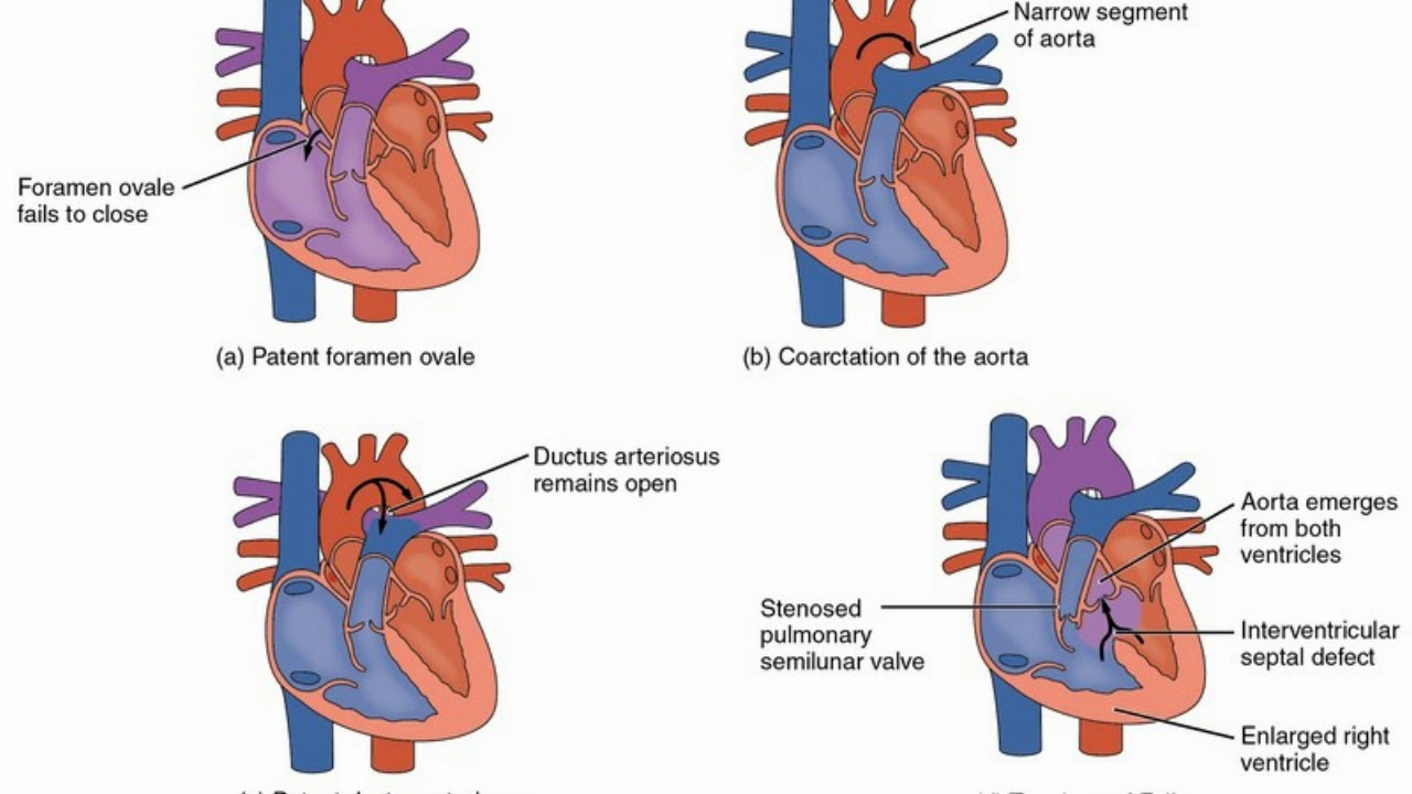
This condition is a sub category of congenital heart defects. Acyanotic heart defects are congenital cardiac malformations that affect the atrial or ventricular walls heart valves or large blood vessels.
 Difference Between Cyanotic And Acyanotic Congenital Heart Defects Youtube
Acyanotic heart disease is a set of heart problems that usually arise prior to or at birth.

Acyanotic heart defect. Common causes include genetic defects e g trisomies maternal infections e g rubella or maternal consumption of drugs or alcohol during pregnancy. Acyanotic heart defects are congenital cardiac malformations that affect the atrial or ventricular walls heart valves or large blood vessels. They do not however hinder the quantity of oxygen or blood that is to be relayed to the tissues.
Common causes include genetic defects e g trisomies maternal infections e g rubella or maternal use of drugs or alcohol during pregnancy. People often retain normal levels of oxyhemoglobin saturation in systemic circulation. An acyanotic heart defect is a class of congenital heart defects in these blood is shunted flows from the left side of the heart to the right side of the heart most often due to a structural defect hole in the interventricular septum.
The intracellular concentrations of these 2 ions appear to be closely correlated but the existence of a relationship between the plasma concentrations of these ions has been controversial. A major function of potassium is to maintain th.
 Magnesium And Potassium Healthy Walker
Surprisingly it also plays a role in nerve impulses muscle movement and blood clotting.

Magnesium and potassium levels. A high potassium level represents a more dangerous condition and can cause significant abnormal heart rhythms 1. That said a low potassium diet is rarely the cause of potassium deficiency or hypokalemia. Both potassium and magnesium are electrolyte minerals meaning they are required for conduction of electrical impulses in the body along with sodium calcium and chloride.
Deficiency is characterized by a blood potassium level below 3 5 mmol per liter 2. Magnesium and potassium are the 2 major intracellular cations. Well magnesium inhibits the romk channels at certain intracellular concentrations.
Sodium rda is about 2300mg. Potassium rda is about 4700mg. Looking at the minerals separately.
If your patient has low magnesium hypomagnesemia then the romk is going to want to waste away the potassium making the patient hypokalemic. Magnesium is necessary for dna and rna production contributes to nerve and muscle function regulates heart rythym regulates blood sugar levels and boosts immune function. Together potassium and magnesium are crucial to heart function and when potassium levels are low magnesium levels are usually low as well.
Magnesium can play a bigrole in helping to regulate the balance of potassium sodium and other electrolytes in the body. Electrolytes play a role in chemical exchanges conduct. Calcium is the most important mineral for tooth and bone density.
This means that until the magnesium is fixed you can t fix the hypokalemia. Low levels of magnesium cause. Excessive alcohol use and intestinal ailments are other possible causes.
Magnesium recommended daily amount is about 400mg. It can also help. High levels of magnesium can cause respiratory depression and cardiac arrest.
Doctor emi s potassium and magnesium citrate is an easy to absorb source of potassium and magnesium which can help normalize muscle function help maintain normal blood pressure levels improve the body s mineral balance and normalize in some cases minor feelings of palpitations that may result from mineral deficiency. The flood gates will remain open. These obviously approximates as the rda doesn t always cut it when it comes to info just using these as a reference.
Potassium and magnesium deficiency can be caused by diuretics and some medications.
Inserting a nasogastric ng tube allows you to directly access a patient s stomach. By inserting an ng tube you are gaining an entry or direct connection to the stomach and its contents.
 Verifying Ng Feeding Tube Placement In Pediatric Patients American Nurse
The nasogastric tube is connected to suction to facilitate decompression by removing stomach contents.

Inserting ng tube. It is very important that the procedure must be done by a trained medical practitioner like doctors or nurses. Patients have an ng tube inserted immediately after any major surgery for approximately 48 72 hours. Simple steps for inserting an ng tube.
Nasogatric ng tubing is a procedure that nurses use for diagnostic and therapeutic purposes. When inserting the nasogastric tube it may be helpful to place your other hand behind the patient s head to keep him or her from pulling back. Gastric decompression is indicated for bowel obstruction and paralytic ileus and when surgery is performed on the stomach or intestine.
Ng tubes can also be used for enteral feeding initially. Therapeutic indications for ng intubation include. You can use ng tubes to drain the stomach take samples and or distribute nutrients and medications.
Nasogastric tube insertion procedure. Nasogastric tubes has varying sizes measured in french 8 10 12 14 16 and 18 fr. By inserting a nasogastric tube you are gaining access to the stomach and its contents.
Inserting the tube is a straightforward process but must be done carefully to minimize the risk of irritation. There are several absolute contraindications for insertion so you should be aware of these. Nasogastric tubes are contraindicated in the presence of severe facial trauma cribriform plate disruption due to the possibility of inserting the tube intracranially.
Creative commons licensing attribution non commercial. An ng tube is intended for short term used to help prevent vomiting after surgery and to keep the patient s stomach empty. Essentially you are inserting a tube from the patients nose into their stomach.
Asking the patient to take sips of water when passing the nasogastric tube through the pharynx into the esophagus and through the esophagus into the stomach can greatly improve the chance of success and reduce gagging. 1 wash hands and prepare materials to be used in the nasogastric tube insertion. Simple steps for inserting an ng tube.
This process is known as nasogastric ng intubation. Creative commons licensing attribution non commercial. Nasogastric ng tubes may be used for feeding or for drainage read your instructions thoroughly as this will dictate the type of tube you need to use.
Additionally the use of cookies provides overall safety while visiting the invicta site. 22340 4 7 out of 5 stars 1 001 99 89 99.
 Invicta Men S 6051 Venom Reserve Black Chronograph Watch For Sale Online Ebay
Invicta pro diver 1773 men s quartz watch 43mm.

Invicta watches mens. In fact both invicta watches for mens and invicta watches for womens come with exceptional designs and high end technology that offers the best value for your. Discover invicta men s quartz watch stainless steel swiss mvt mod 33970. Invicta is one of the most recognizable swiss watch company since 1837.
Invicta men s watch aviator chrono gold tone and black dial steel bracelet 17205. It looks elegant and sophisticated in the cool black and silver colors and has an impressive 40mm stainless steel case with a mineral dial window. Was 595 00 88 off.
The modest price of invicta watch australia has nothing to do with the brand s outstanding designs and durability. We use cookies to better understand how you use the invicta site so that we can personalize content and advertising to improve your user experience. For more information on cookies or to opt out of this website s use of cookies please click here.
Invicta pro diver 12819 women s quartz watch 40mm. Invicta i force 19656 men s round analog chronograph black polyurethane watch. 299 00 130 00.
Discover the official invicta stores to shop the most famous exclusive watches online for men women. The invicta men s 8926ob is a stunning round watch that features a corrugated unidirectional bezel and luminous hands markers. Invicta 24768 mens speedway white mop dial chronograph steel bracelet dive watch.
299 00 105 00. Invicta is synonymous to high quality yet affordable swiss watches. Like most invicta watches this one also features an original analog japanese quartz movement.
Invicta men s pro diver scuba 50mm gold tone stainless steel and silicone chronograph quartz watch black gold model. Invicta 8928ob pro diver unisex wrist watch stainless steel automatic blue dial by invicta. Invicta pro diver 1772 men s quartz watch 43mm.
Was 995 00 92 off. Invicta 21865 pro diver men s wrist watch stainless steel automatic blue dial by invicta. Invicta speedway 9223 men s quartz watch 39 5mm.
299 00 105 00.
Diverticulosis is the formation of abnormal pouches diverticula in the bowel wall. Usually the pain is severe and comes on suddenly but it can also be mild and become worse over several days.
 Diagnosis And Management Of Acute Diverticulitis American Family Physician
Acute pain r t outward protruding out of the mucosa and sub mucosa in the gastrointestinal tract characterized by the client complaining of abdominal pain the client appears nervous.
Diverticulitis care plan. Pain related to physical injuring agents bacterial invasion causing edema irritability spasm of colon. Nursing care plans for diverticulitis nursing care plan 1. Diverticulitis nursing care plan.
Nursing care plan for diverticulitis. Peristomal are related to sensitivity to the materials used. Nursing diagnosis for diverticulitis.
These home remedies may help you feel. Fever chills are signs of infection and possibly early peritonitis. Diverticulitis is a disease of the intestines in which small often painful pouches form.
Their doctor or health care provider. Acute pain related to presence and inflammation of diverticula as evidenced by pain score of 10 out of 10 verbalization of right upper quadrant abdominal pain and cramping guarding sign on the abdomen abdominal rigidity and restlessness. Risk for impaired tissue integrity.
Diverticulitis is inflammation or infection of these pouches. Diverticulosis is common and frequently has no symptoms however when many diverticula are present the normal. It is the main duty of the nurse to provide the patient with all the measures that are needed to make the colon rest especially during the acute pain and disturbance which happens when the bacteria present in the food or otherwise causes the inflammation.
The most common sign on examination is tenderness in the lower left side of the abdomen. These pouches may become infected and can be a medical crisis. Maintain npo status during initial phase of antibiotic treatment to kill infection and help bowel rest as symptoms decrease advance diet to clear liquids and then increase fiber slowly.
6 27 pm diverticulitis nursing care plan 5 comments. Nursing care plan for diverticulitis. Constipation r t narrowing of the colon secondary to thickening of the muscle segment and structure characterized by the client saying bloating of the abdomen.
These conditions are known as diverticular disease. The most common symptom of diverticulitis is abdominal pain. Peristomal skin and tissue remain intact.
Diverticulitis occur when fecal material becomes lodged in these pouches causing inflammation and bacterial invasion. Maintain the integrity of the peristomal tissue.
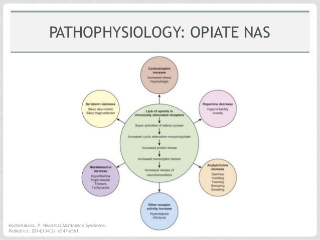
Neonatal abstinence syndrome neonatal abstinence syndrome nas or neonatal withdrawal syndrome nws occurs when in utero addictive substances are suddenly discontinued due to birth. Or they may start as late as 5 to 10 days after birth.
 Neonatal Abstinence Syndrome
Withdrawal from licit or illicit substances is becoming more common among neonates in both developed and developing countries.
Pathophysiology of neonatal abstinence syndrome. Nas is most often caused when a woman takes drugs called opioids during pregnancy. Key points about neonatal abstinence syndrome. Neonatal abstinence syndrome also called nas is a group of conditions caused when a baby withdraws from certain drugs he s exposed to in the womb before birth.
Neonatal abstinence syndrome happens when babies are exposed to drugs in the womb before birth. The most common substances include alcohol nicotine and rapidly increasing opioids. Neonatal abstinence syndrome nas is often a multisystem disorder that frequently involves the cns gastrointestinal gi system autonomic system and respiratory system.
Nas is diagnosed every 25 minutes in the united states. Neonatal abstinence syndrome nas is a collection of symptoms experienced by babies whose mother has abused certain drugs during pregnancy. It is a multisystemic disorder resulting from chronic in utero exposure and.
During pregnancy substance abuse is on the rise especially opioids both prescribed and illicit resulting in a hidden epidemic of neonatal abstinence syndrome nas. Neonatal abstinence syndrome nas is a result of the sudden discontinuation of fetal exposure to substances that were used or abused by the mother during pregnancy. Symptoms of withdrawal may begin as soon as 24 to 48 hours after birth.
Withdrawal from licit or illicit substances is becoming more common among neonates in both developed and developing countries. It can occur in pregnancies where the mother takes drugs such as heroin alcohol methadone or even marijuana. Babies can then be affected or go through drug withdrawal after birth.
However rat models show differences between neonatal and adult withdrawal process. Neonatal abstinence syndrome nas is a result of the sudden discontinuation of fetal exposure to substances that were used or abused by the mother during pregnancy. Neonatal abstinence syndrome is influenced by many factors including maternal fetal placental pharmacokinetics neurotransmitter dysregulation genetic and epigenetic factors.
In reality it can occur either before or after the baby is born. The pathophysiology underlying nas has not been fully understood.
Sounds intermediate between bronchial and vesicular breath sounds. These bronchovesicular breath sounds are normal if heard between 1st and 2nd intercoastal spaces.
These sounds can be heard using a stethoscope or simply when breathing.

Bronchovesicular breath sounds definition. Observed anteriorly and posteriorly in between scapulae. Farlex partner medical dictionary farlex 2012. Bronchovesicular sounds means sounds that are present between bronchial and vesicular breath sounds.
In contrast vesicular breath sounds are soft low pitched predominantly inspiratory and appreciated especially well at the posterior lung bases. Bronchovesicular breath sounds definition these are a mixture of bronchial breath sound during expiration and vesicular breath sound during inspiration with no gap in between the inspiration and expiration. These are normal sounds in the mid chest area or in the posterior chest between the scapula.
The inspiratory component predominates and is generated by turbulent airflow within the lobar and segmental bronchi whereas the expiratory component is due to flow within the larger airways. These breath sounds include crackles wheezes stridor and pleural rubsl these are explained in the essentials of lung sounds lessons. Lung sounds also called breath sounds can be heard across the anterior and posterior chest walls.
If heard in other areas of the lung bronchial sounds are abnormal. They reflect a mixture of the pitch of the bronchial breath sounds heard near the trachea and the alveoli with the vesicular sound. Bronchovesicular sounds can be heard during inspiration and expiration and have a mid range pitch and intensity.
Vesicular breath sounds are the sounds heard during auscultation of the chest of a healthy person listen to the audio recording below. Breath sounds come from the lungs when you breathe in and out. They can be abnormal but are normal when heard between the 1st and 2nd intercostal spaces anteriorly and posteriorly between scapulae.
They have an i e ratio of 1 1. Breath sounds can be normal or abnormal.
You have already completed the quiz before. You can participate in quizlet for rhythm identification and cardiac rhythm interpretation.
 Practice 12 Lead
Hence you can not start it again.

Ecg practice test strips. We suggest you practice with these prior to taking the post test. The following rhythm strips are for your practice. Practice with over 200 electrocardiogram strips as well as factual guidelines for rapid and efficient 12 lead ecg interpretation.
Improve your ecg knowledge with our free ecg quiz. Ecg interpretation experience takes a long time to acquire in the field. Stay with me here.
Here are the 2 ways to classify ekg rhythms. Just strips names learn with flashcards games and more for free. Use these ekg practice tests to help you become proficient in your rapid rhythm identification.
Put your knowledge to the test with over 200 ecg strips. All strips are six second strips unless otherwise indicated rhythm strip 1 ecg criteria. This ekg practice test is designed to help you learn to recoginze all of the ekg rhythms that you will encounter during emergencies and during the aha acls provider course.
There are 2 ways of classifying ekg rhythms making it a whole lot easier to interpret any rhythm presented to you be it in clinical setting in class acls or in an ekg rhythm test. I have even included a rhythm practice strips with answers and explanation. Select your difficulty level beginner intermediate advanced or random and get started now.
Basic procedures adult part i adult part ii pediatric pulmonary hypertension. Ekg practice drills ekg reference guide ecg graded quiz ekg intro course ecg tutor ecg monitor drill ecg monitor quiz 12 lead ecg ecg 12 lead interpretation tutor español ekg lessons. American heart association aha acls advanced cardiac life support acls ecg rhythm strips pretest practice review question answers quiz pdf.
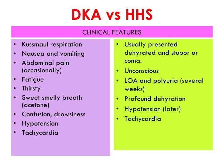
Dka and 15 for hhs 1 2 dka is the most common cause of death in children and adolescents with type 1 diabetes and accounts for half of all deaths in diabetic patients 24 years of age 6 the cause of death in patients with dka and hhs rarely results from the metabolic complications of hyper glycemia or metabolic acidosis but. Thus ketones are produced and the patient may present with a mix of both dka and hhs.
 Dka Vs Hhs Suraya
Diabetic ketoacidosis dka and hyperosmolar hyperglycemic state hhs are the two most serious metabolic complications of diabetes mellitus dm.

Dka and hhs. 12 the diagnostic criteria for hhs is defined as hyperglycemia often with blood. Diabetic ketoacidosis dka and hyperglycemic hyperosmolar state hhs are acute metabolic complications of diabetes mellitus that can occur in patients with both type 1 and 2 diabetes mellitus. Timely diagnosis comprehensive clinical and biochemical evaluation and effective management is key to the successful resolution of dka and hhs.
Dka presents in patients with type 1 dm t1dm while hhs is more commonly seen in patients with type 2 dm t2dm. Diabetic ketoacidosis and hyperosmolar hyperglycemic syndrome hhs are life threatening emergencies that occur in patients with type 1 diabetes and type 2 diabetes 1 2 3 4 diabetic ketoacidosis is defined by a triad of hyperglycemia or a diagnosis of diabetes metabolic acidosis and ketonemia 1 2 5 6 7 hhs is defined by severe hyperglycemia high serum osmolality and dehydration 4 8 the presentation of each of these diabetic emergencies often overlaps 3 8 early diagnosis and. Hyperosmolar hyperglycemic state hhs 1 2 severe hyperglycemia hyperosmolality and dehydration in the absence of significant ketoacidosis.
In severe cases of hhs the insulin resistance is so great that the body does begin to break down fatty acids. Dka is characterized by hyperglycemia ketone body formation and metabolic acidosis. 6 often these patients are obese and extremely insulin resistant.
Diabetic ketoacidosis dka 1 2 uncontrolled hyperglycemia metabolic acidosis and increased total body ketones. These disorders can occur in both type 1 and type 2 dm. Dka vs hhs dka means diabetic ketoacidosis and hhs means hyperosmolar hyperglycemic syndrome both dka and hhs are the two complications of diabetes mellitus.
Diabetic ketoacidosis dka and hyperglycemic hyperosmolar state hhs are complications that present in the acute phase of diabetes mellitus dm. They are associated with increased risk of mortality 1 2. Diabetic ketoacidosis dka and hyperosmolar hyperglycemic syndrome hhs are two acute complications of diabetes that can result in increased morbidity and mortality if not efficiently and effectively treated.
When comparing the two hhs has a higher mortality rate. Though there are many differences between dka and hhs the basic problem is associated with insulin deficiency.